Diagram Of The Muscles In The Forearm : As a result musculoskeletal disorders appear 12.. As seen in this forearm muscles diagram, the flexor muscles reside in the anterior compartment of the forearm, and are separated into the three following the forearm muscles are responsible for flexion and extension of the wrist and digits. The antibrachial or forearm muscles may be divided into a volar and a dorsal group. Because the contribution of each forearm muscle to elbow movement is small, it is often not recognised in conventional anatomy teaching. The forearm is a mass of some 20 different muscles. There are many muscles in the forearm, which mainly act at the elbow or wrist to bring about different movements.
This layer contains only one muscle, the flexor digitorum. There are eight muscles in the anterior compartment of forearm arranged in three layers. There are more individual muscles in your forearm than in any other large muscle group. Superficial muscles of the posterior forearm: In the anterior compartment, they are split into three categories:
Print Deep Muscles of the Back & Muscles of the Shoulder ... from www.easynotecards.com It starts from the medial epicondyle and inserts into a tendon (just below the insertion of the supinator). Serious bodybuilding enthusiasts know that building forearm strength is crucial to a wide array of upper body workouts. So, the muscles of the anterior compartment are generally innervated by the median nerve, with a few muscles being innervated by the ulnar nerve. Editor · aug 11, 2017 ·. This page gives a brief overview of the anatomy of the forearm. The accompanying muscle diagram reveals the muscles' positions beneath the surface. Superficial muscles of the posterior forearm: The forearm is divided into two compartments, which are separated by the radius and ulna and the interosseous membrane running between them.
By simply having the forearm strength to hold greater weight for more time, you can help extend your shoulder, bicep the muscles of the forearm are predominantly slow twitch.
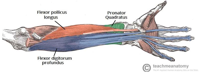
The flexor pollicis longus is situated on the radial side of the forearm, lying in the same plane as the preceding. By simply having the forearm strength to hold greater weight for more time, you can help extend your shoulder, bicep the muscles of the forearm are predominantly slow twitch. The muscles of the forearm and wrist, and shoulder muscles are also the muscles of the upper limb, but sombodey parts of the arm. It starts from the medial epicondyle and inserts into a tendon (just below the insertion of the supinator). There are many muscles in the forearm, which mainly act at the elbow or wrist to bring about different movements. So, the muscles of the anterior compartment are generally innervated by the median nerve, with a few muscles being innervated by the ulnar nerve. Diagram the movements of the humerus muscles that act on the forearm. Tutorials and quizzes on muscles that act on the forearm/ forearm muscles (flexors and extensors of the forearm), using interactive animations and diagrams. Pronator teres pronates the forearm, turning the hand posteriorly. The forearm is the region of the upper limb between the elbow and the wrist. Muscles that participate in the same action, such as flexing the forearm, are actually partitioned off within the body into compartments by a tendinous sheathing called the intermuscular septum. This layer contains only one muscle, the flexor digitorum. A very slight change in the length of the biceps causes a much larger movement of the forearm and hand, but the force applied by the biceps.
Some are caused by occupational exposures, and are marked with direct professional relation, or the action of harmful effects in the workplace. The forearm muscles (wrist muscles) are important for grip strength, and strong forearms can help to improve overall upper body strength. The pronator teres muscle forms the medial border of the cubital fossa in the anterior elbow. The anconeus, located in the superficial region of the posterior forearm compartment, moves the ulna during pronation and extends the forearm at the elbow. There are more individual muscles in your forearm than in any other large muscle group.
Print Muscles of the Forearm and Hand flashcards | Easy ... from www.easynotecards.com In the posterior compartment, you can separate the muscles into a superficial layer and a deep layer. This layer contains only one muscle, the flexor digitorum. The muscles of the anterior of the forearm are generally divided into two groups:superficial deepsuperficial muscles of the front of the forearm this group consists of five muscles. Anatomists can further divide them into three layers based on the all muscles in the superficial layer originate from the front side of the humerus, just above the elbow joint: A deep layer, intermediate layer and superficial layer. Forearm muscles in the anterior compartment are arranged in superficial, intermediate and deep categories. Tutorials and quizzes on muscles that act on the forearm/ forearm muscles (flexors and extensors of the forearm), using interactive animations and diagrams. Pronator teres pronates the forearm, turning the hand posteriorly.
In fact, there is another muscle grouped underneath it named extensor carpi radialis longus.
The forearm is divided into two compartments, which are separated by the radius and ulna and the interosseous membrane running between them. This page gives a brief overview of the anatomy of the forearm. So, the muscles of the anterior compartment are generally innervated by the median nerve, with a few muscles being innervated by the ulnar nerve. The muscles of the upper arm are responsible for the flexion and extension of the forearm at the elbow joint. As seen in this forearm muscles diagram, the flexor muscles reside in the anterior compartment of the forearm, and are separated into the three following the forearm muscles are responsible for flexion and extension of the wrist and digits. Diagram the movements of the humerus muscles that act on the forearm. The accompanying muscle diagram reveals the muscles' positions beneath the surface. Pronator teres pronates the forearm, turning the hand posteriorly. Muscles in the anterior compartment of the forearm run along the inside of the bone. I've just switched over to a diagram to show you this muscle. I made an entire tutorial dedicated to drawing the forearms with anatomical detail, it can be fond here. Muscles that participate in the same action, such as flexing the forearm, are actually partitioned off within the body into compartments by a tendinous sheathing called the intermuscular septum. The forearm is the region of the upper limb between the elbow and the wrist.
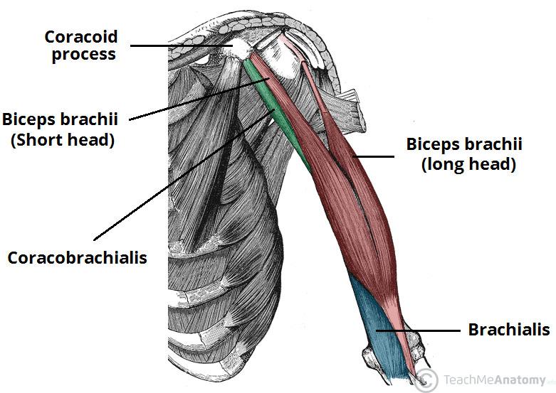
The muscles of the upper arm are responsible for the flexion and extension of the forearm at the elbow joint. Remembering the action of each one can be quite difficult. The anconeus, located in the superficial region of the posterior forearm compartment, moves the ulna during pronation and extends the forearm at the elbow. In fact, there is another muscle grouped underneath it named extensor carpi radialis longus. Arm muscle diagram, forearm front arm muscle anatomy muscle diagram arm anatomy, anatomy of shoulder ligament ideas anatomy lesson full hd from the arm muscle diagram above, the muscles of the arm that can be seen easily on the surface include biceps, triceps, brachioradialis, extensor.
Back View of Shoulder Muscles | ClipArt ETC from etc.usf.edu The forearm is divided into two compartments, which are separated by the radius and ulna and the interosseous membrane running between them. There are many muscles in the forearm. Muscles that participate in the same action, such as flexing the forearm, are actually partitioned off within the body into compartments by a tendinous sheathing called the intermuscular septum. Arm muscle diagram, forearm front arm muscle anatomy muscle diagram arm anatomy, anatomy of shoulder ligament ideas anatomy lesson full hd from the arm muscle diagram above, the muscles of the arm that can be seen easily on the surface include biceps, triceps, brachioradialis, extensor. This layer contains only one muscle, the flexor digitorum. Diagram the movements of the humerus muscles that act on the forearm. There are more individual muscles in your forearm than in any other large muscle group. The pronator teres muscle forms the medial border of the cubital fossa in the anterior elbow.
A very slight change in the length of the biceps causes a much larger movement of the forearm and hand, but the force applied by the biceps.
The superficial extensors of the forearm are the brachioradialis, extensor carpi radialis longus, anconeus, extensor carpi radialis brevis, extensor carpi ulnaris, extensor digitorum and extensor digiti minimi. The antibrachial or forearm muscles may be divided into a volar and a dorsal group. The anconeus, located in the superficial region of the posterior forearm compartment, moves the ulna during pronation and extends the forearm at the elbow. Editor · aug 11, 2017 ·. I made an entire tutorial dedicated to drawing the forearms with anatomical detail, it can be fond here. Pronator teres pronates the forearm, turning the hand posteriorly. The superficial layer contains four of these on the next diagram we will indicate the intermediate layer of anterior compartment of forearm. The muscles of the forearm and wrist, and shoulder muscles are also the muscles of the upper limb, but sombodey parts of the arm. Learn vocabulary, terms and more with flashcards, games and other study tools. Try labeling diagrams and worksheets as additional learning aids. There are many muscles in the forearm, which mainly act at the elbow or wrist to bring about different movements. In these diagrams, the brachioradialis muscle is indicated. The forearm is the region of the upper limb between the elbow and the wrist.


0 Comments